




ADVANCED MRI DIAGNOSTICS
Detailed Imaging Without Ionising Radiation with Siemens MAGNETOM Essenza
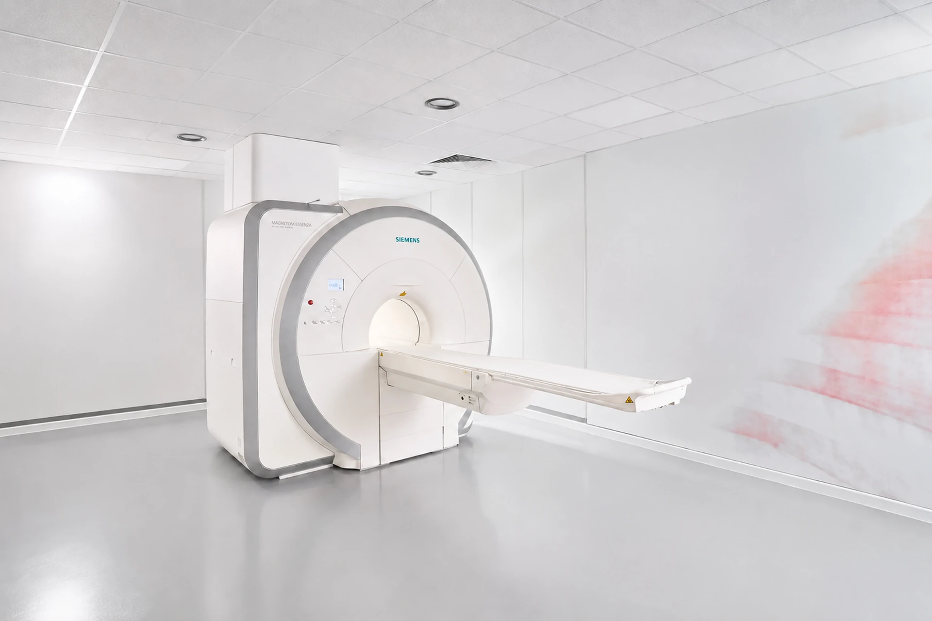
At PHI Megamed+, magnetic resonance imaging is performed using a 1.5 T MRI system designed to provide detailed imaging of soft tissues, an efficient examination process and greater patient comfort.
WHAT IS MAGNETIC RESONANCE IMAGING?
Exceptionally Detailed Imaging of Soft Tissues
Magnetic resonance imaging (MRI) is a non-invasive diagnostic imaging method that uses a strong magnetic field, radio waves and computer processing to create detailed images of the internal structures of the body. Unlike X-rays and CT scans, MRI does not use ionising radiation.
Siemens MAGNETOM Essenza provides imaging with a 1.5 T magnetic field and Tim technology, allowing flexible combinations of receiver elements and protocols for a wide range of clinical examinations—from the head and spine to the joints, abdomen and pelvis.
- Excellent contrast between different types of soft tissue
- Multiplanar imaging without changing the patient’s position
- No X-rays or ionising radiation
SYSTEM ADVANTAGES
Technology Designed for Detailed and Consistent Imaging
MAGNETOM Essenza combines a homogeneous 1.5 T magnetic field with Tim receiver technology and clinical protocols for precise imaging of different anatomical regions.
Soft-tissue detail
The high contrast of MRI images helps visualise the brain, spinal cord, nerves, muscles, ligaments and internal organs.
No ionising radiation
The method uses a magnetic field and radiofrequency energy rather than X-rays, which is an important advantage when repeated follow-up examinations are required.
Tim technology
The combination of multiple receiver elements allows flexible coverage of the anatomical region and parallel imaging in multiple directions.
Greater comfort
The ultra-short magnet and lightweight receiver coils are designed to facilitate more comfortable positioning and a better patient experience.
More “head-out” examinations
For certain anatomical regions, the short magnet design allows the patient’s head to remain outside the magnet for most of the examination.
Advanced sequences
Different MRI sequences and fat-suppression techniques provide additional information according to the clinical indication.
WHEN IS MRI USED?
A wide range of diagnostic examinations
Magnetic resonance imaging is particularly valuable when detailed imaging of soft tissues, the nervous system, joints and internal organs is required.
Important: The specific protocol, anatomical region and need for a contrast agent are determined by the physician based on the referral diagnosis, medical history and clinical indication.
SIEMENS MRI TECHNOLOGY
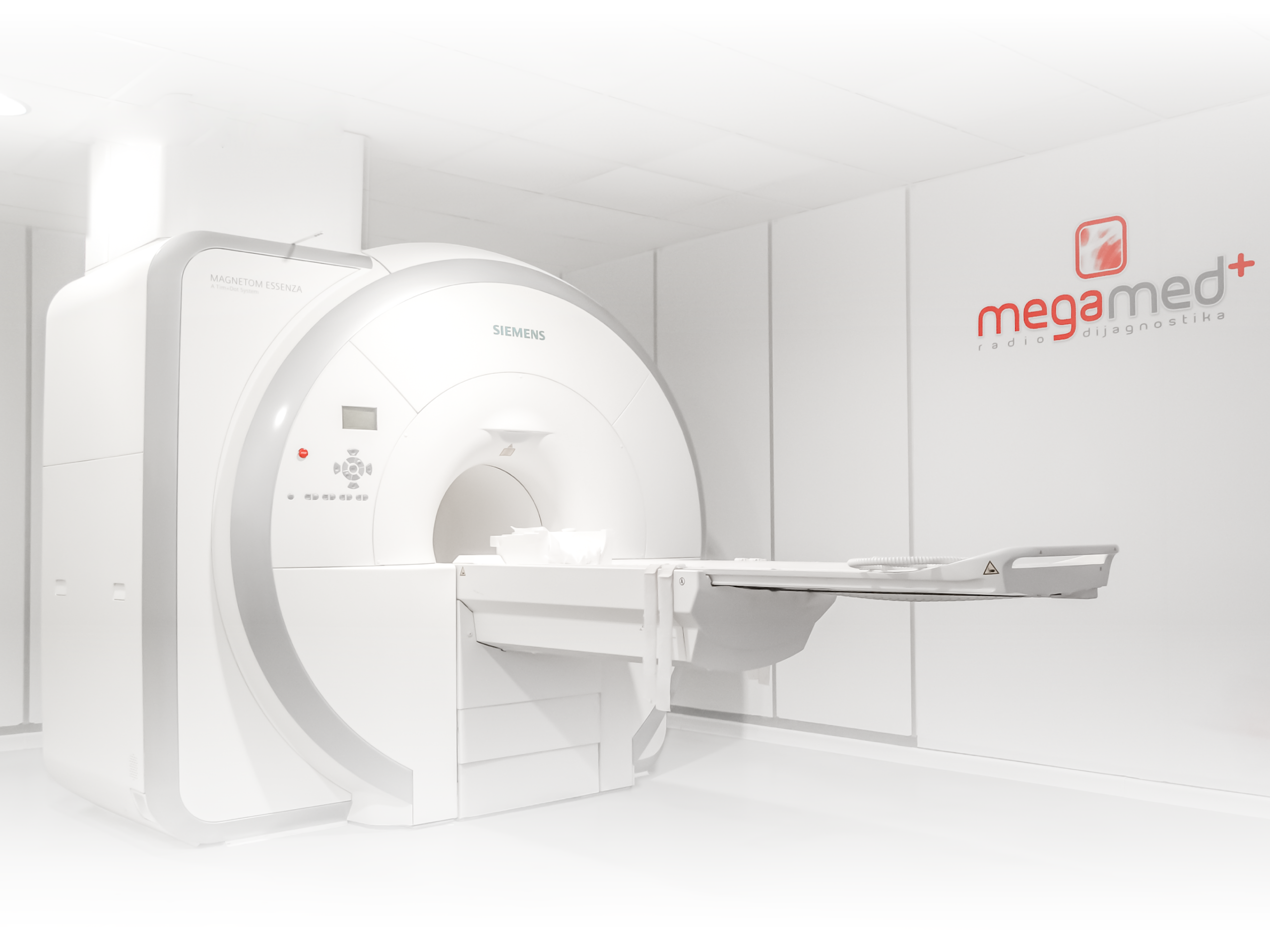
MAGNETOM Essenza – 1.5 T and Tim Technology
MAGNETOM Essenza is designed as a compact and reliable 1.5 T MRI system. Tim (Total Imaging Matrix) technology enables flexible combinations of 25 receiver elements using up to four coils, providing extended field of view and parallel imaging.
At PHI Megamed+, this technology is complemented by careful safety screening, clear instructions, proper positioning and a protocol tailored to the specific clinical indication.
*The specifications listed above are based on the manufacturer’s official documentation. Certain applications, functions and coils may depend on the configuration and software options of the specific system.
BEFORE YOUR EXAMINATION
How should you prepare?
Safety screening is mandatory before every MRI examination. When you make your appointment, you will receive instructions based on the area being examined and whether contrast may be used.
Bring your medical records
Bring your referral, previous imaging, medical reports and documentation for any implanted medical devices or implants.
Report implants and metal
Tell us if you have a pacemaker, cochlear implant, neurostimulator, pump, clip, dentures, metal fragment or have undergone previous surgery.
Remove metal objects
Before entering the examination room, you will need to remove jewellery, watches, phones, cards, hearing aids, and clothing or accessories containing metal components.
Share relevant health information
Tell us if you are pregnant or breastfeeding, have kidney disease, allergies or claustrophobia, or have previously experienced a reaction to contrast.
Never assume that an implant is MRI-safe. A medical device with an unknown safety status must not enter the MRI environment until its model has been identified and the conditions for safe scanning have been confirmed.
FREQUENTLY ASKED QUESTIONS
What do patients most often want to know?
MRI does not use X-rays or ionising radiation. Images are produced using a strong magnetic field, radio waves and computer processing.
The examination is non-invasive and generally painless. You will need to lie still, and the scanner produces loud rhythmic sounds, so appropriate hearing protection is provided.
The duration depends on the anatomical region being examined, the number of sequences and whether contrast is required. You will receive more specific information about your examination when you make your appointment.
Only if the exact model has been identified and is labelled MR Safe or MR Conditional, and all of the manufacturer’s conditions can be met. A device with an unknown safety status is treated as unsafe for MRI until proven otherwise.
No. Gadolinium-based contrast agent is used only when necessary for the specific clinical indication. Before contrast is administered, your medical history, kidney function, allergies and pregnancy status are assessed.
Inform the team when you make your appointment. You will receive an explanation of how the examination is performed, and for certain anatomical regions, the short magnet design allows a larger part of the body to remain outside the magnet. Any medication, if necessary, is prescribed by the physician.
Precise diagnosis begins with the right technology and careful preparation
Contact PHI Megamed+ for information, safety screening, preparation instructions and to schedule your MRI examination.
This information is provided for general informational purposes only and does not replace a medical examination, medical advice, MRI safety screening or an individual assessment by a radiologist.